- Home
- About Us
- Meet Our Team
- Patient Forms
- Hearing Evaluations And Testing
- Hearing Aid Fitting and Repair
- Custom Earmolds And Ear Plugs
- Tinnitus Management
- Types Of Hearing Aids
- Hearing Aids We Carry
- Caring For Your Hearing Aids
- Types of Hearing Loss
- How The Ear Works
- Dizziness and Balance
- Your Balance System
- Vestibular Rehabilitation
- Balance Testing
- Aftercare
- Blog
- Contact us
About Us
Hearing
Dizziness and Balance
TESTIMONIALS
See What Our Happy Clients
Are Saying
How the balance system works
The space orientation and equilibrium system work by using reference points and objects surrounding us; these reference points are controlled by some specialized receptors that tell our brain our position in space, and how we are moving in relation to those objects.
There are three different kinds of specialized balance receptors:
- Vestibular System receptors: located in the inner ear; inform the brain of the position and movement of the head in relation to space.
- Visual-oculomotor system receptors: located in the retina (eye), inform the brain about position and movement of eyes within the visual field.
- Musculoskeletal / somatosensorial system receptors: inform the brain about the movement of different parts of the body in relation to each other and the ground.

All three systems coordinate and partially correlate their information, some of these systems generate overlapping information, and this allows the brain to regulate, and even repair, itself. This regulation and repair is needed to keep space orientation and equilibrium in difficult situations such as boat trips or rollercoasters; these regulatory mechanisms even allow the brain to compensate for pathologies affecting one or more of the “balance systems”. This compensation can be achieved spontaneously or with the help of external stimulus such as Vestibular rehabilitation.
The first step in the process of recovery is to identify the source of the dizziness; an effective diagnostic process is key in determining the available and adequate treatment options.
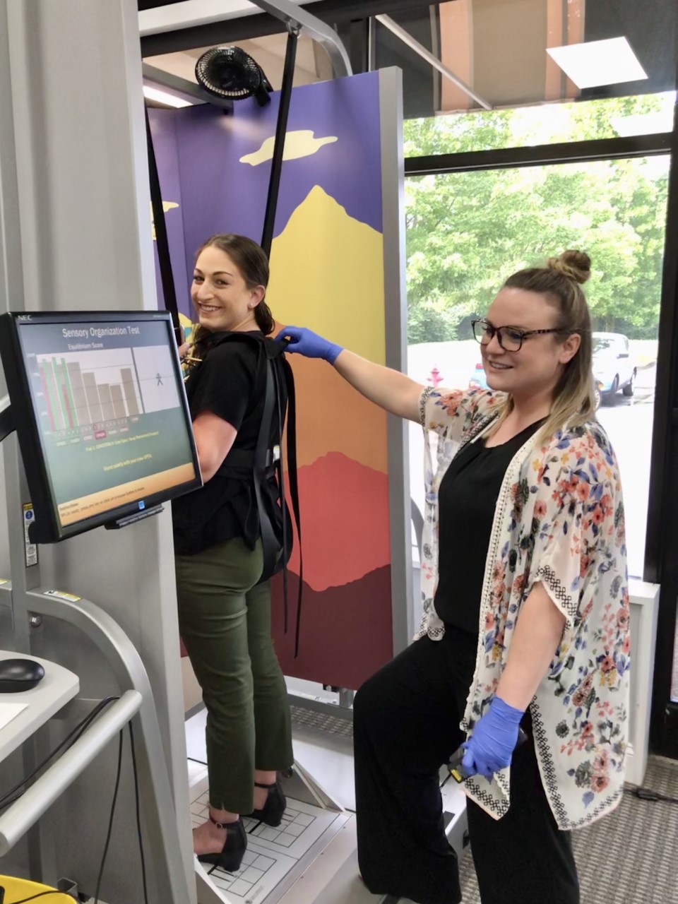
Computerized Dynamic Posturography
Historically, the quantitative measurement of equilibrium was something rarely done due to the complexity of the systems involved and the lack of reliable methods to do so. However, now we have several tools we can use to quantify the balance or equilibrium, the Computerized Dynamic Posturography (CDP) is probably the most use done due to its accessibility and multiple applications.
CDP is performed with the patient standing on a mobile platform with sensors that detect patient’s sway and a visual surround that can move. Six different conditions in which the support platform and visual surround will change their status, are presented to the patient and their reactions measured to obtain a balance score and an estimate of the contribution of the different systems that contribute to balance.

Vestibular Tests
The vestibular tests allow us to assess the ocular movements that maintain the images of our surroundings stable on the retina, this is the basis of balance and space orientation.
Videonystagmography (VNG)
Videonystagmography is an assessment that allows us to register and analyze the ocular movements in different situations and in response to different stimulus. VNG assessment consists of the following tests:
Spontaneous Nystagmus
We search for the presence of absence of involuntary spontaneous eye movements called nystagmus, nystagmus has a characteristic rhythm and frequency and can be originated by vestibular injuries (most frequently) or the failure of the cerebral mechanisms that control the ocular movements.

Oculomotor Testing
This test assess the oculomotor system and its controllers (brain and cerebellum), it is performed by presenting a visual target that moves slow or fast depending on the studied mechanism and the ocular movements are then recorded and analyzed.
Positional Testing
Positional testing searches for the presence of nystagmus and symptoms of dizziness triggered by specific positions of the head and body and most of the times are caused by vestibular alterations. The eye movements and their relationship to the position that elicits them points to a specific origin and affected structure, each of these with a different treatment.
Rotary Chair
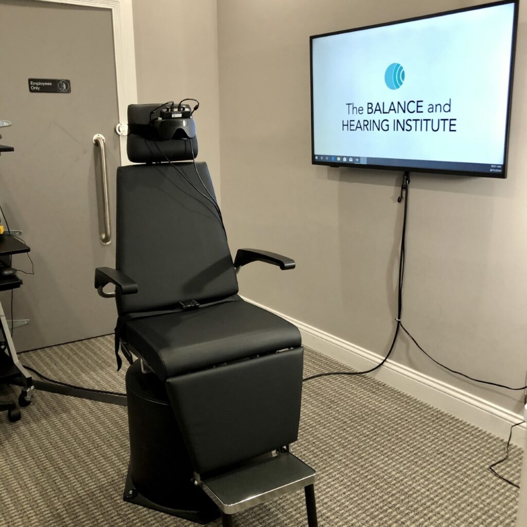
Step rotation test allows us to measure the function and symmetry of the vestibular nerves and the Semicircular canals (they act like the sensors for movement). During this test the chair will rotate slowly for one minute to one side while eye movements are recorded, then we will repeat the process rotating the chair to the other side. The chair will move slowly, smoothly and it is usually well tolerated.
-OR-
Caloric Testing
Caloric testing consists on the assessment of the vestibular nerves by individually stimulating them and recording the ocular movements provoked by this stimulation. It is done by irrigating each ear with warm (50*C) and cool (24*C) air.

Electrophysiological Tests
Auditory Brainstem Response (ABR) and Electrocochleography (ECoG)
ABR and ECOG are non-invasive tests to assess the status of the Auditory Nerve and the Cochlea, they are performed with the patient comfortably laying down and relaxed, a pair of electrodes is adhered to the forehead and earphones like foam inserts are placed in both ears, then sound is presented to each ear for a brief period of time, patient does not need to respond to these sounds.
Vestibular Evoked Myogenic Potentials (cVEMP and oVEMP)
VEMPs assess the function of the saccule and utricle (two small structures located within the inner ear) which, due to connections of the vestibular nerve, elicit a muscle contraction in response to noise. This test is performed by placing electrodes on the skin of the neck and/or around the eyes and presenting noise on each ear.

There are a variety of balance tests – contact us to learn more about what could be right for you!
Request an Appointment

